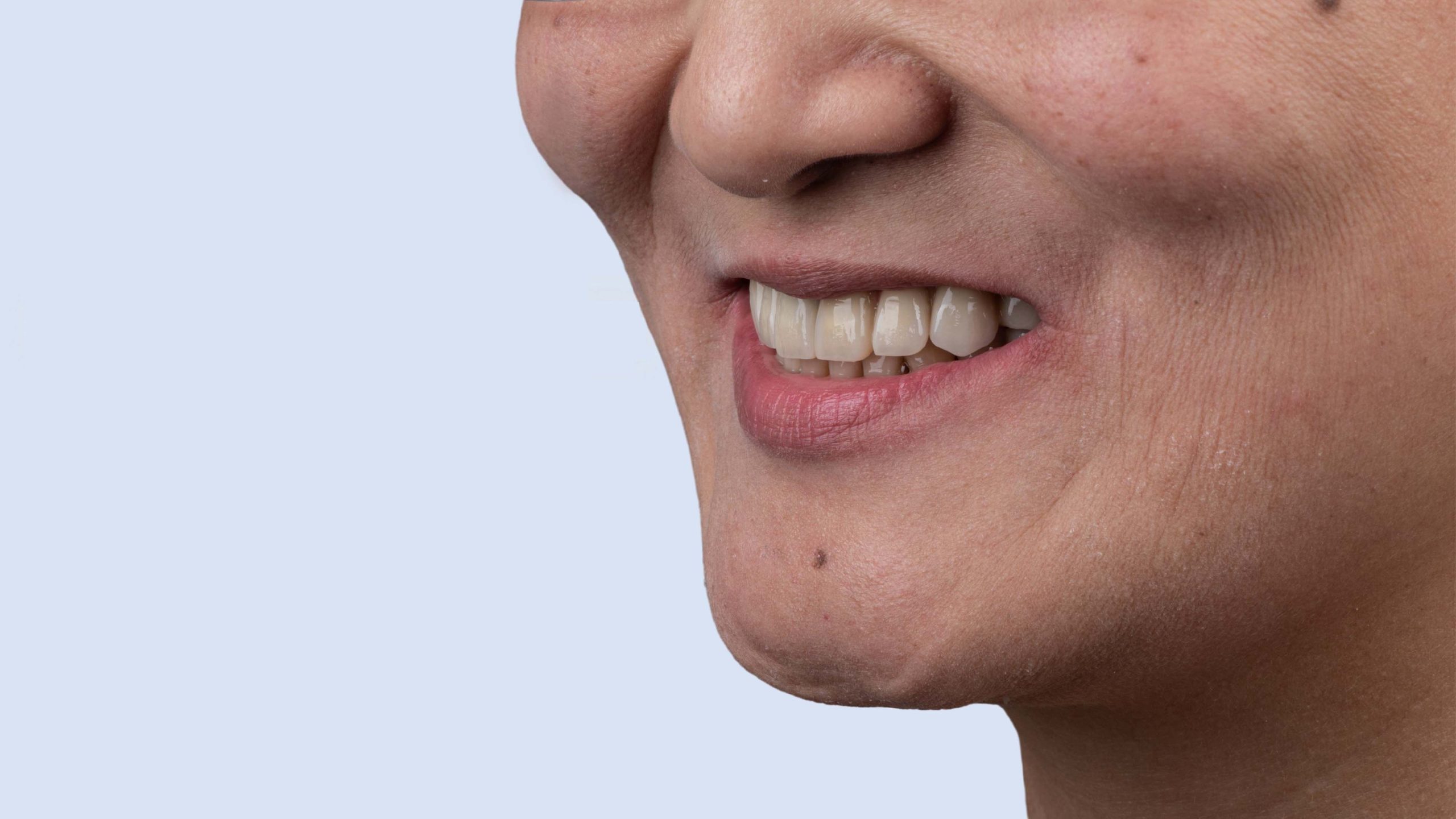
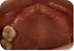
A 44 years old female patient presented with complaints of loose and missing teeth, seeking implant treatment at the hospital. Upon examination, she was found to have complex tooth loss in the upper and lower jaws, with compromised occlusal relationships.
Treatment Plan:
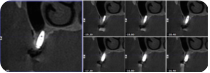
Following preoperative CBCT evaluation, the digital implant treatment plan is as follows:
Establish the jaw relation
Digitize bone augmentation
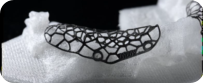
Customize titanium mesh
Digital implantation
Immediate restoration
Final restoration
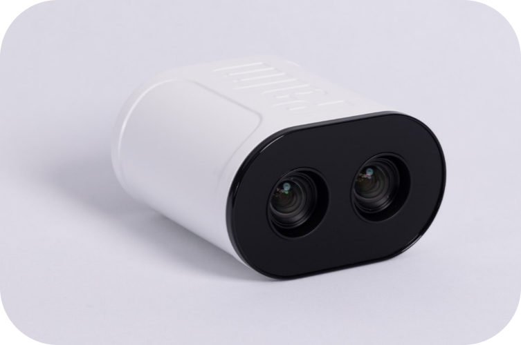
Utilizing PALM Dental Photogrammetry Scanner
The Palm features a high-speed, high-resolution professional infrared camera designed to swiftly and accurately detect the micro Palmark installed on the abutment, facilitating precise digital impression-taking.
Its precision reaches 5 microns, ensuring reliability independent of the technician’s skill and experience in impression-taking. The micro Palmark is compact and lightweight, designed for easy oral installation, and compatible with multiple implant brands.
Compared to traditional methods, it offers higher clinical efficiency, simpler procedures, and increased patient comfort.
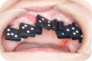
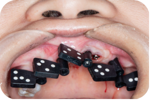
1. Utilize the oral scanning device to capture the patient’s occlusal relationship and soft tissue data while using Palmcap.
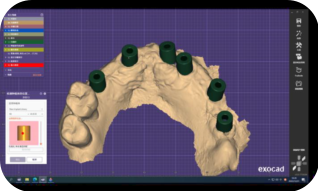
2. Utilize Palm to capture the implant positions.
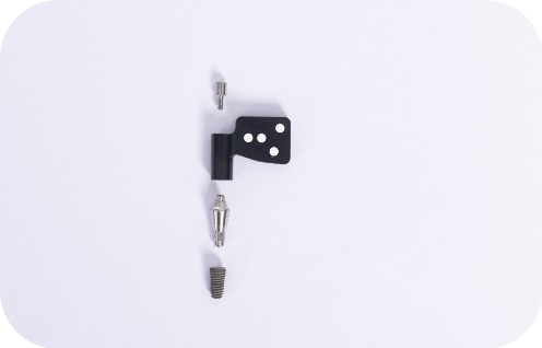
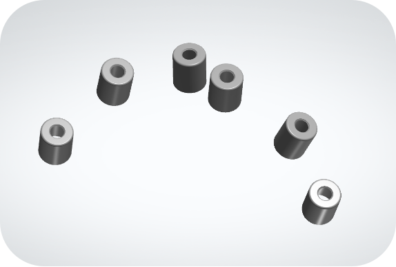
1) Install Palmark
2) Locate Palmark
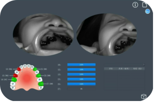
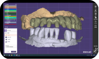
3) Align implant positions data with soft tissue data.
3. Align implant positions data with soft tissue data.Export files in STL format, and use software like exocad to match the data and design bridges.
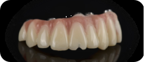
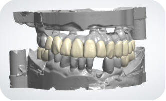
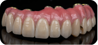
4.The final prosthesis
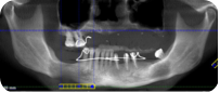
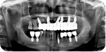
Panoramic radiograph with prosthesis
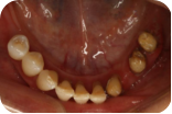
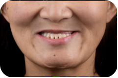
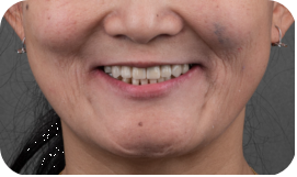
Comparison of anterior and posterior dental restoration
The case was presented by Dr. Huang Yuanding, Chief Physician at the Affiliated Stomatological Hospital of Chongqing Medical University.