The patient, a 67-year-old male, presented with progressive mandibular tooth loss and residual roots in the mouth. The treatment involved half-mouth edentulous jaw guide plate implantation. Post-operation, Palm dental photogrammetry scanner facilitated efficient immediate repair.
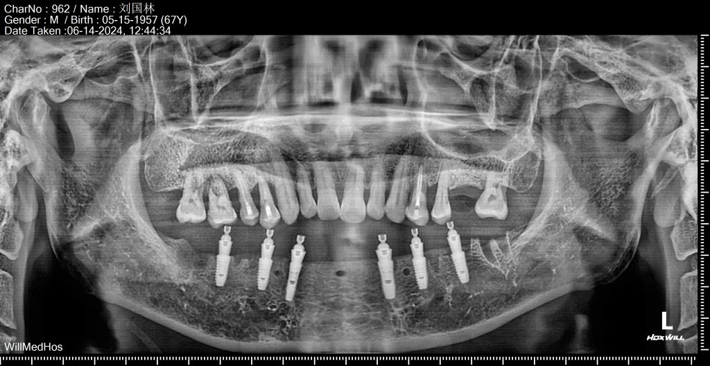
1. Cone Bean Computed Tomography
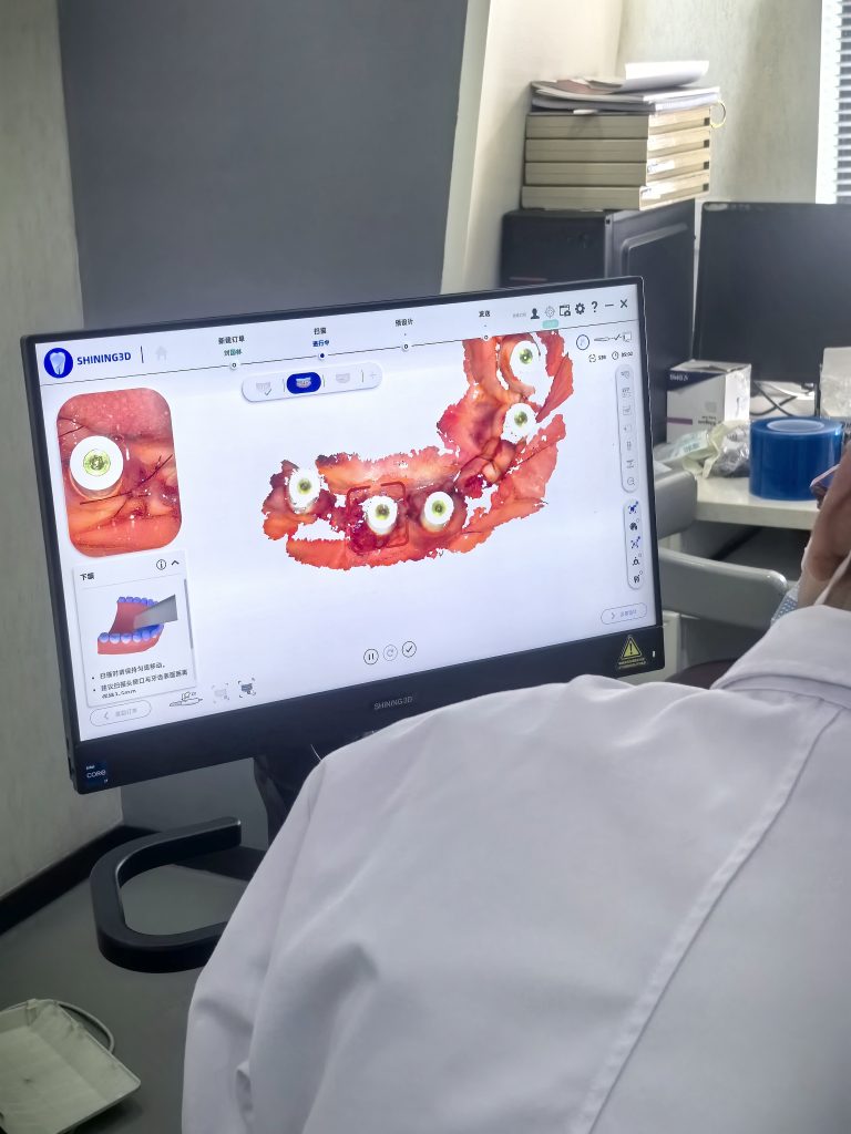
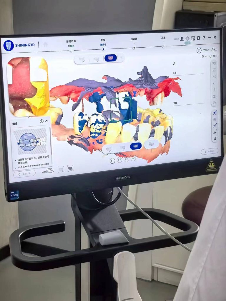
2. Using oral scanner to capture soft tissue data:
1)Install Palmcap
2)Oral scanning
3)Soft tissue data captured
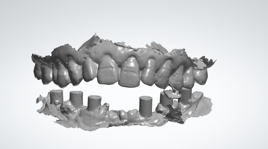
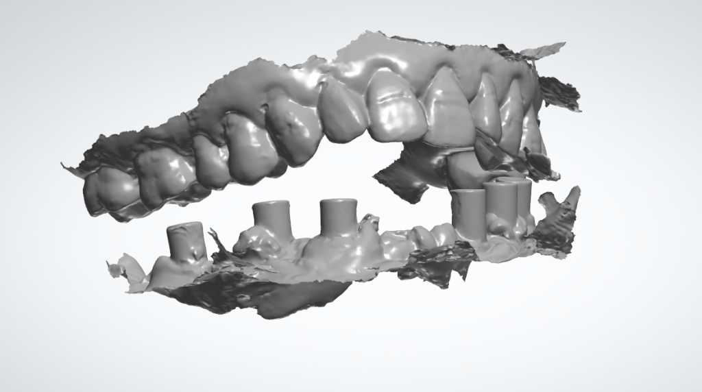
3. Obtaining the patient’s occlusal relationship:
1) Obtaining the occlusion relationship
2) Data on occlusal relationship
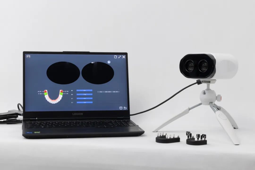
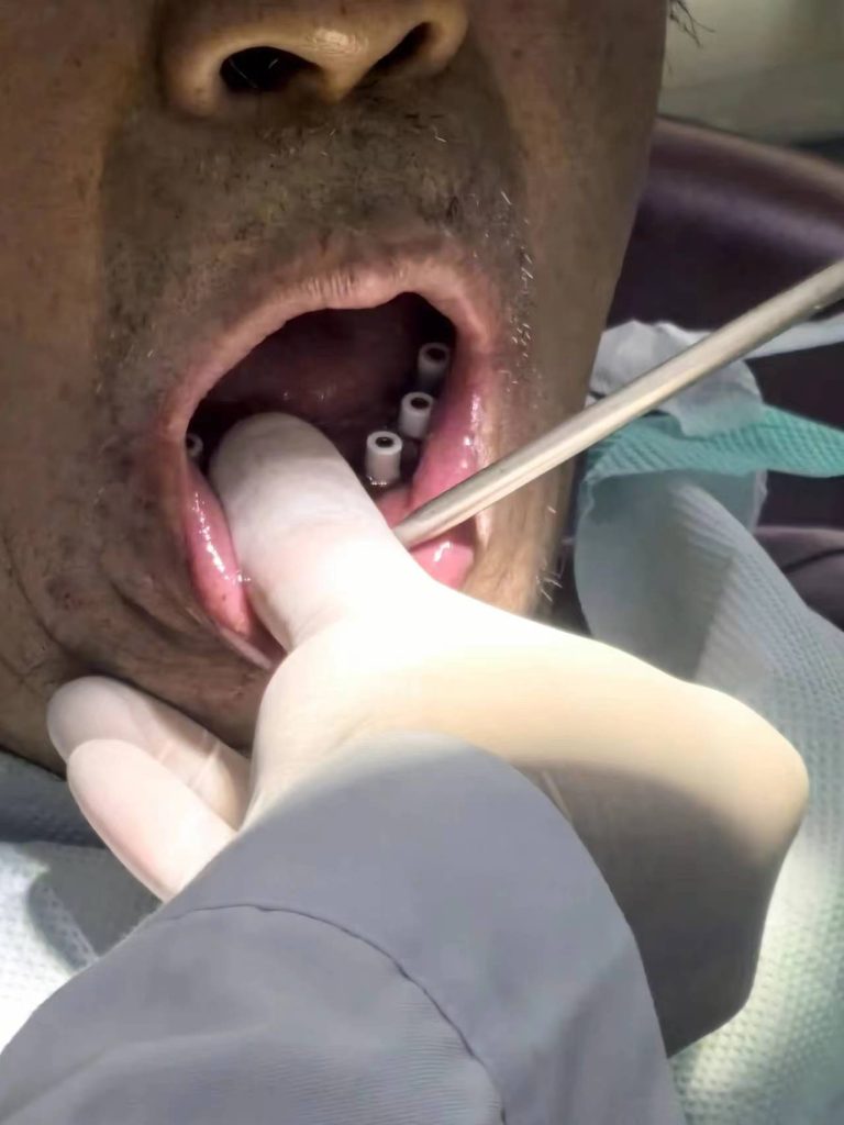
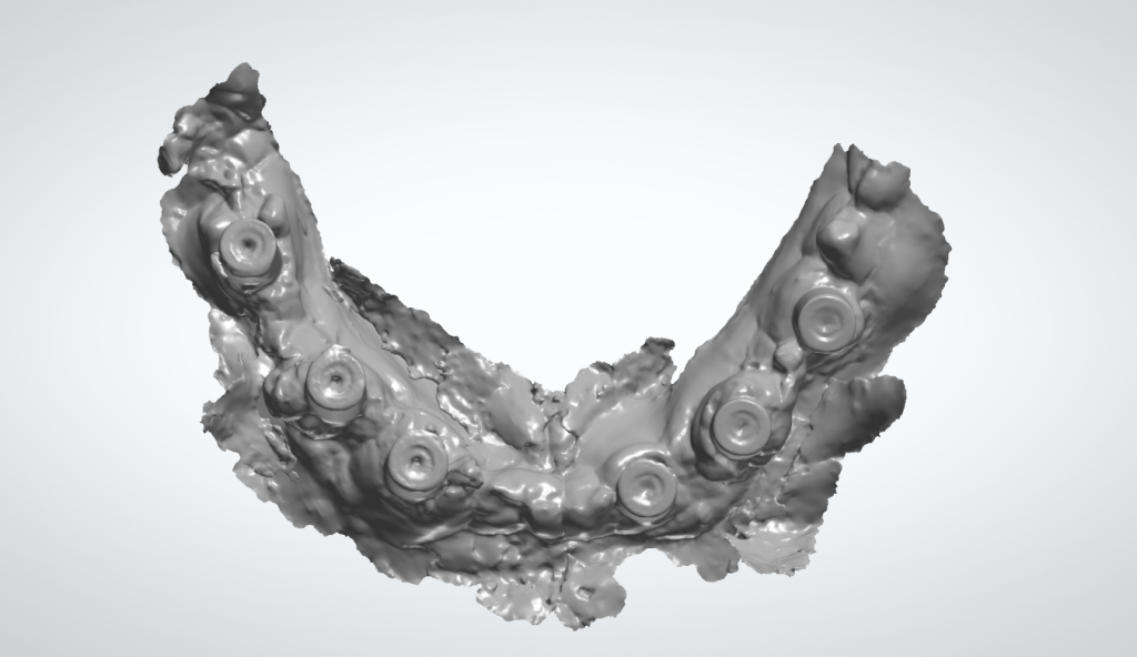
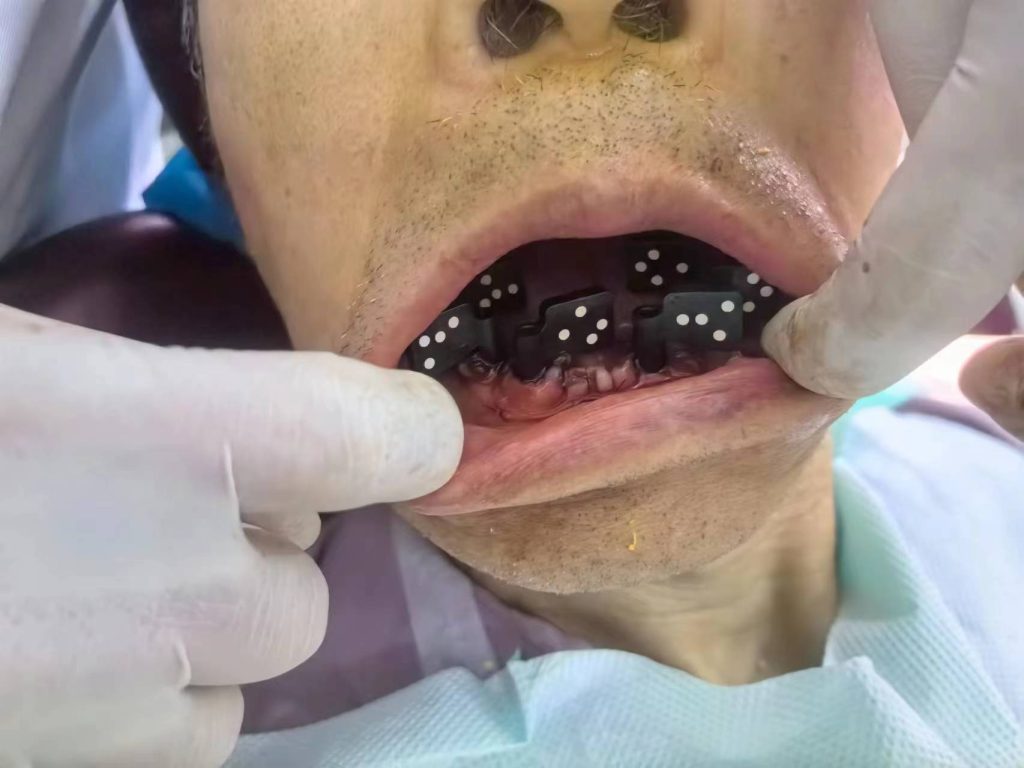
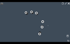
4. Using Palm dental photogrammetry scanner capture detailed data of the abutment:
1)Install Palmark.The smaller and more comfortable Palmark, designed to be convenient for installation and adaptable to various implant systems.
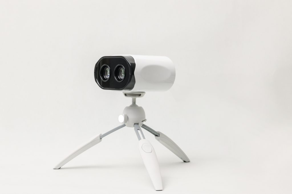
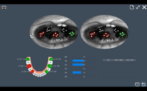
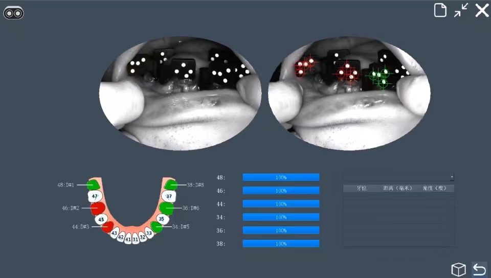
2)Using Palm dental photogrammetry scanner. Palm swiftly and precisely captures the spatial position of the implant abutment. It is compact, lightweight, user-friendly, portable, and convenient to grasp.
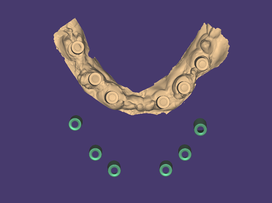
3)Spatial positioning data of the implant abutment. The data can be exported to third-party software in STL format.
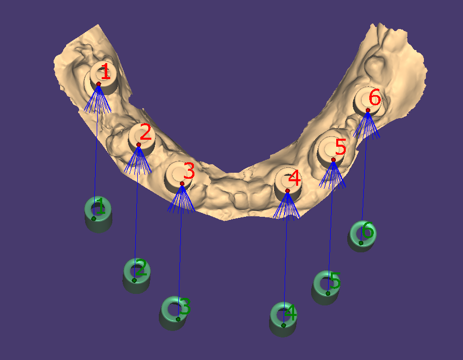
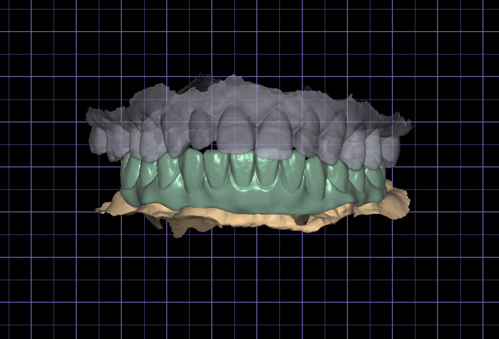
5. Creating temporary prosthesis:
1)Align the spatial location data of the implant abutment with the soft tissue data.
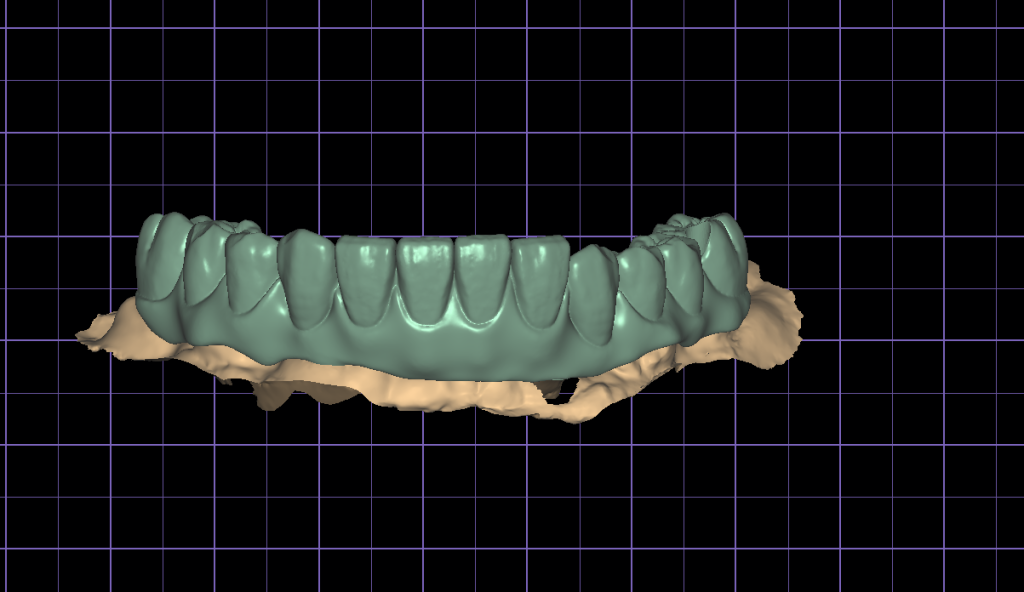
2) Design temporary prosthesis
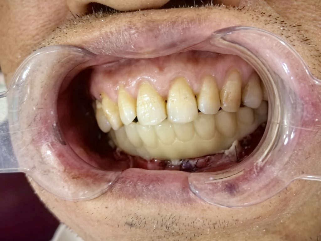
6. Temporary prosthesis
Case Summary
In this case, Palm dental photogrammetry scanner was used to achieve immediate repair, enabling simple, efficient, and comfortable digital impression-taking. This method demonstrates clinical applicability, ensuring successful postoperative temporary prosthesis placement.
Moreover, compared to traditional methods, Palm dental photogrammetry scanner accurately and swiftly captures the implant abutment’s spatial position in the mouth, reducing chairtime and markedly enhancing the doctor-patient experience.