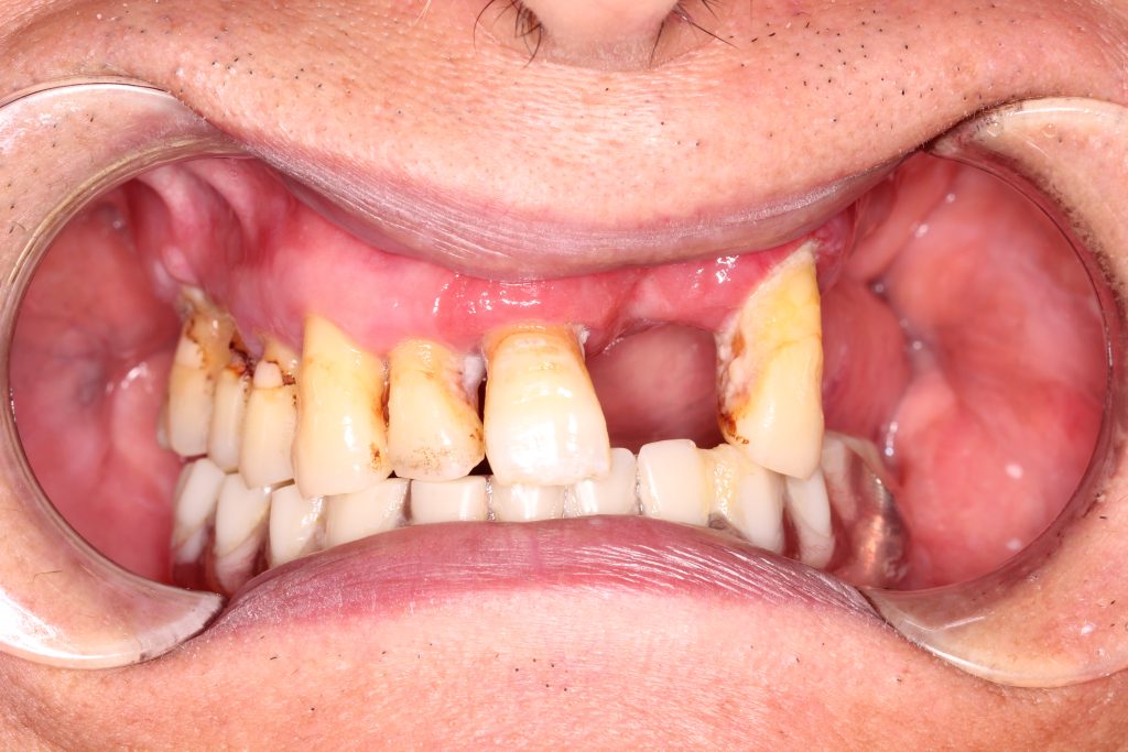
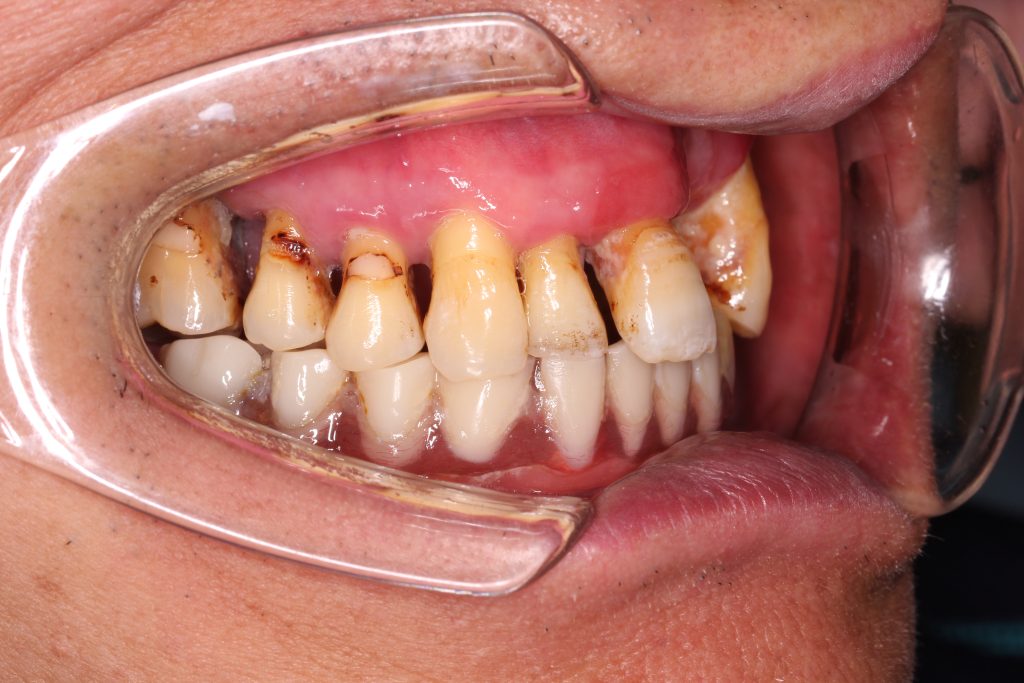
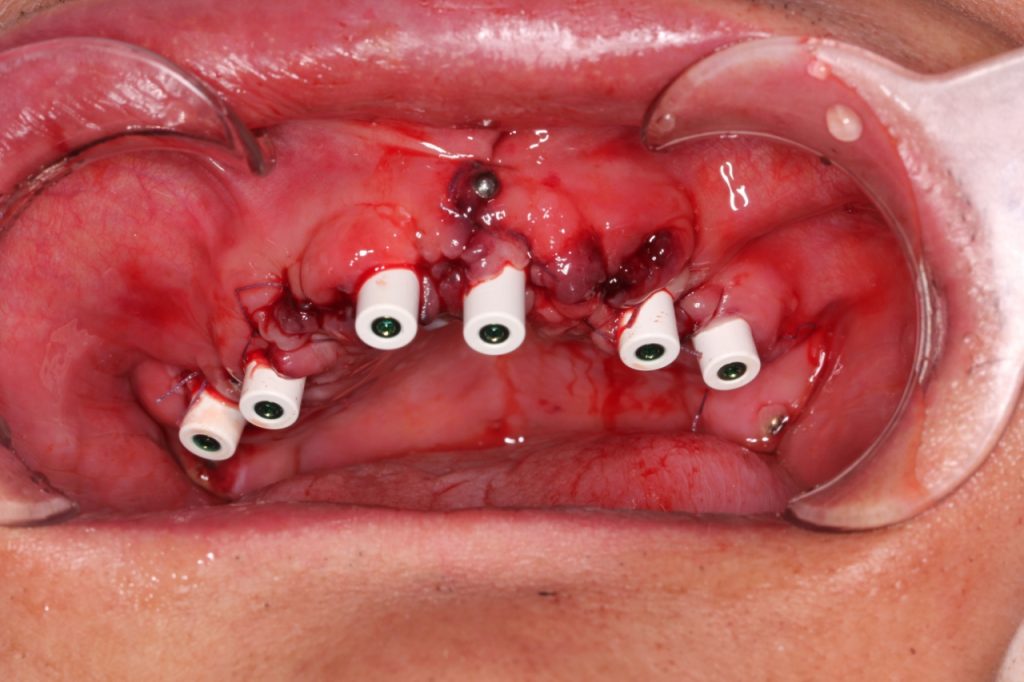
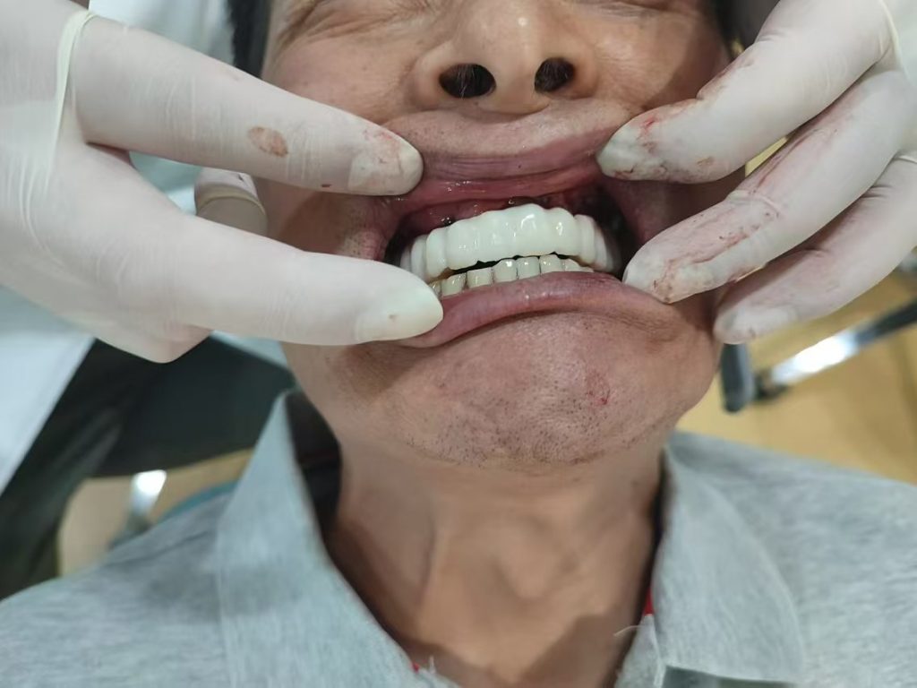
The patient in this case is a 60-year-old male with missing and partially loose maxillary teeth, requiring implants. The treatment plan involves removing the loose teeth and immediately implanting and repairing the upper jaw. After the implants are placed, a Palm oral implant digital impression locator will be used to complete the immediate repair.
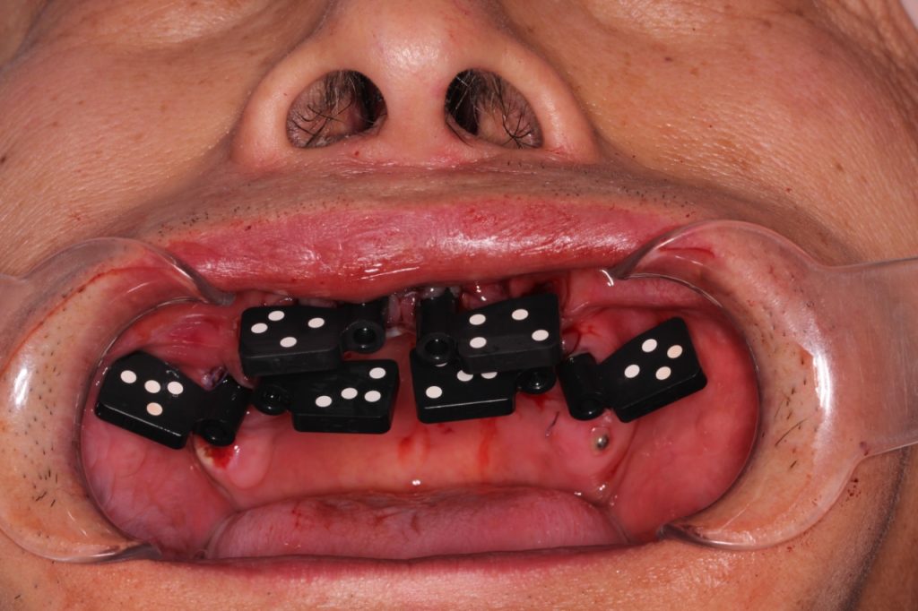
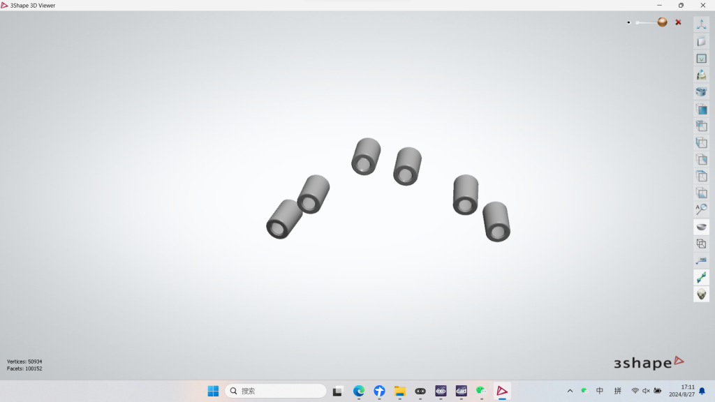
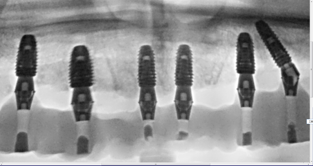
1. Using the Palm dental photogrammetry scanner to capture detailed data of the abutment.
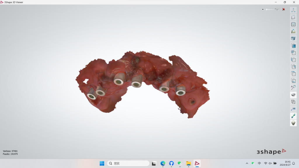
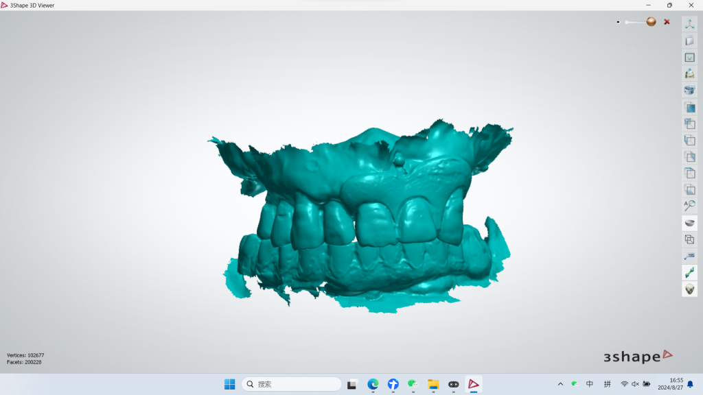
2. Using an oral scanner to capture soft tissue data and obtain the patient’s occlusal relationship.
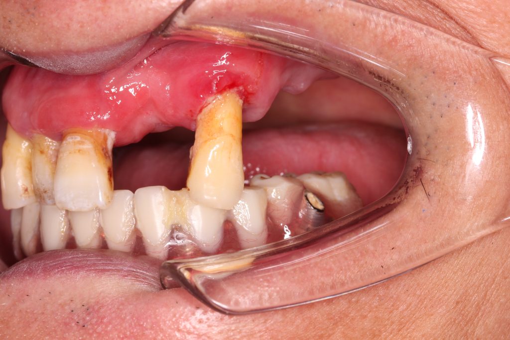
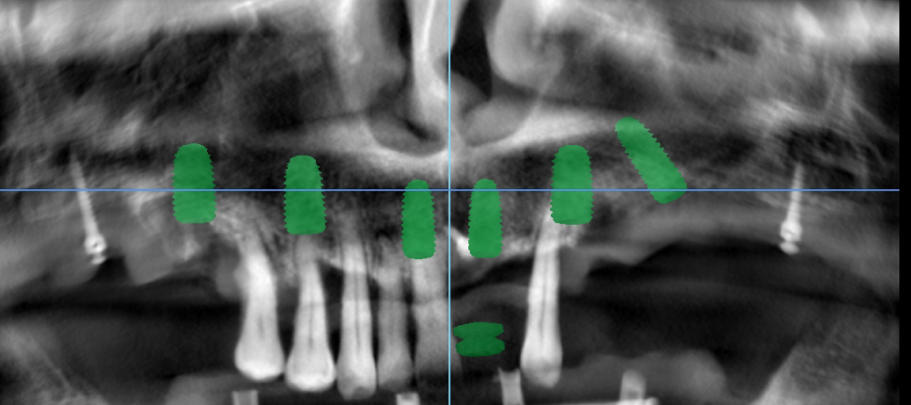
There are seven natural teeth in the patient’s upper jaw, and three bone pins were implanted preoperatively. The patient’s occlusal data was scanned using an oral scanner, which also captured the implanted bone pins in the upper carina along with the occlusal data.
After the patient is ready for implant removal, the implant’s spatial position data is scanned using Palm extraoral scanning. Next, a Palmark is placed inside the patient’s mouth, and the patient’s epithelial mucosa data is captured using oral scanning, which also scans the bone nails along with the implant.
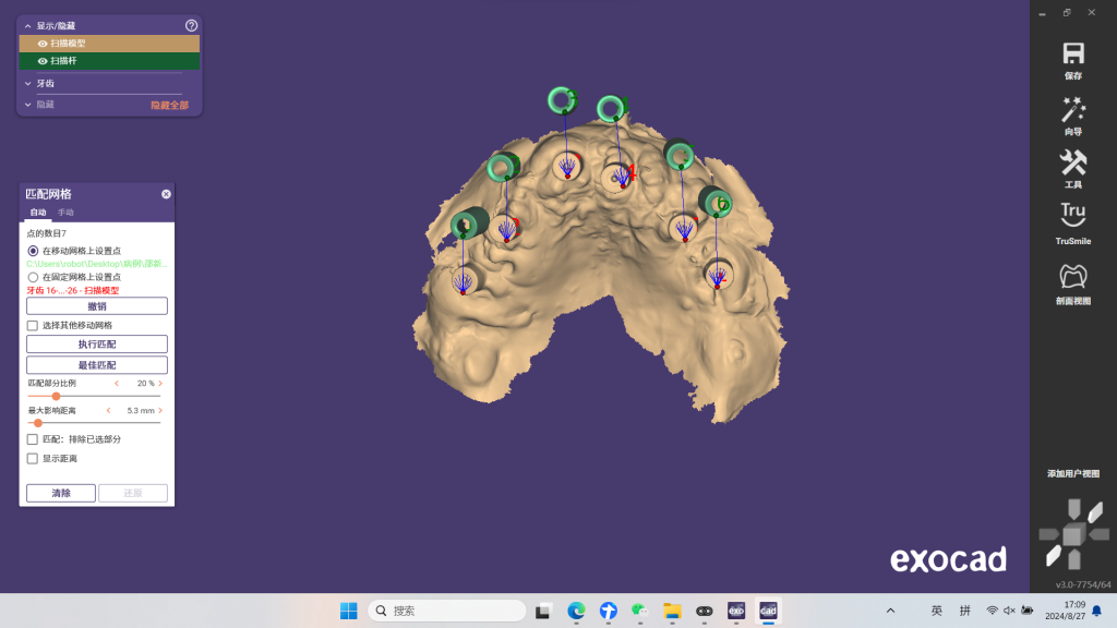
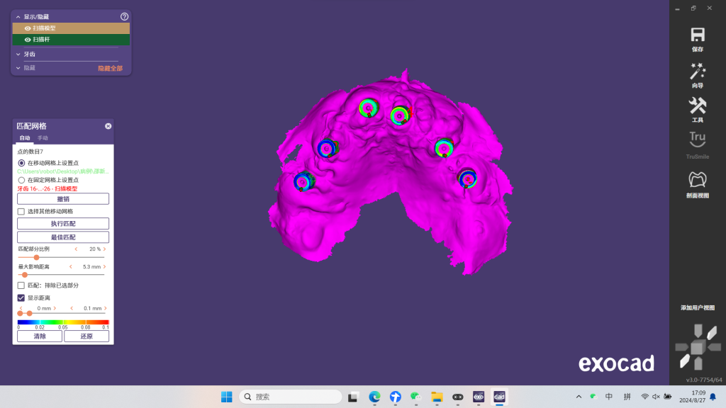
3. Align the spatial location data of the implant abutment with the soft tissue data.
During the third-party software design process, the patient’s occlusion was determined by matching the preoperative maxillary data to the postoperative maxillary data using three bone pins.
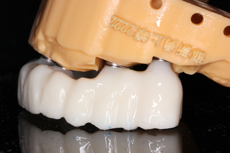
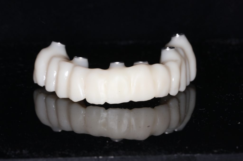
4. Design and create temporary prosthesis
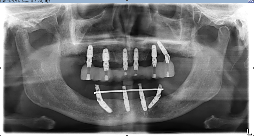
5. Panoramic radiograph with prosthesis
Case Summary: The PALM Dental Photogrammetry Scanner was used for this restoration, enabling a fully digital process with fast and accurate chairside data collection. This approach shortened chairside time, facilitated postoperative wound protection, and ensured the temporary restoration fit well after surgery. Compared to traditional mold-taking, this method also improved patient comfort and postoperative satisfaction.